From clinical needs to patient therapy by addressing the tissue microenvironment its heterogeneity and cell to cell crosstalk and the development of novel technologies and biotherapies.
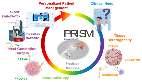
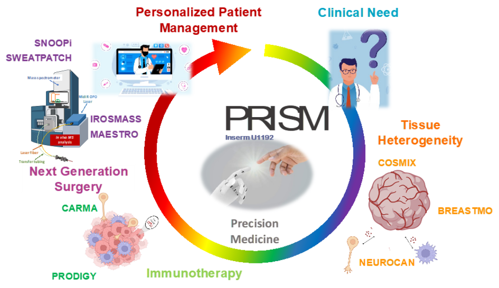
PRISM project will offer a well focus project from clinical needs to therapy. This will include using COMIX tools tumor microenvironment investigation by taking into account the role nerve for tumor progression and Evs for tumor cross-talk. The aims is to identify new markers for immunotherapy enhanced by DDR inhibitors in conjunction with next generation surgery. PRISM will offer to the scientific community and the society novel possibilities to detect BC cancer at early stages, but also novel therapies based on new instruments associated with new biotherapies thanks to fundamental and translational approaches. What PRISM is bringing is the possibility for the future to have a complete autonomous instrument to perform diagnosis, prognosis, surgery and complementary treatment to all solid tumors.
Technological innovations Pillar
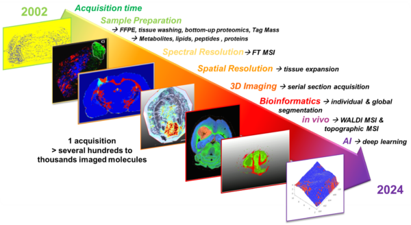
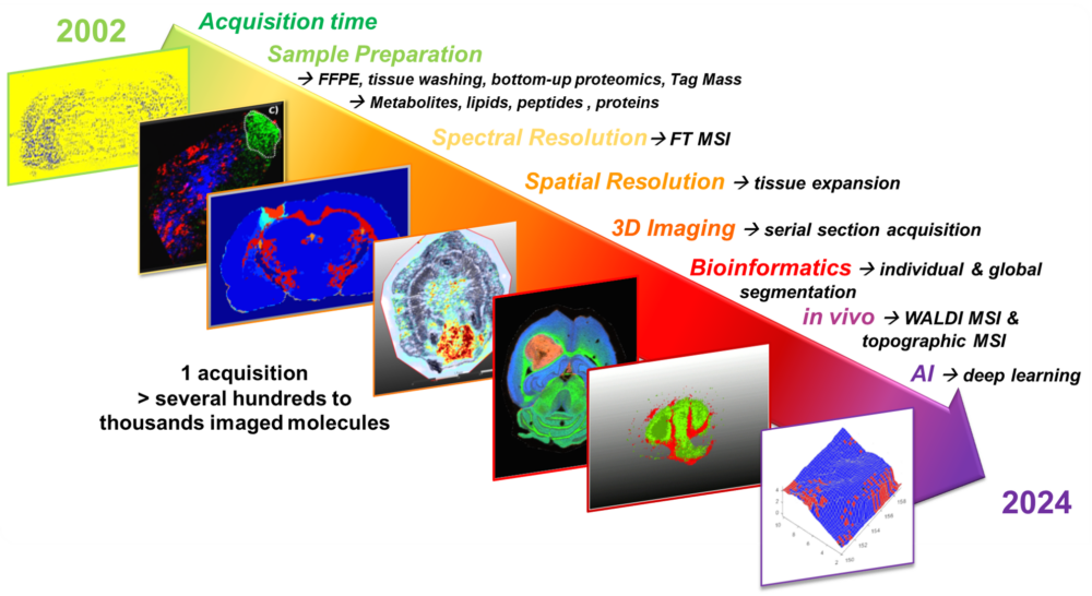
The Technological Innovation axis – (Coordinated by Pr. I. Fournier) arose from the MALDI Imaging Team (MIT) created in 2004 after Prof. I. Fournier obtained a French National Starting Grant (ACI young researchers) based on the research that was conducted since 2002 on MALDI MS Imaging (MALDI MSI) 1, 2. Since 2004, this research was organized around i) technological developments and ii) clinical applications transversally to the second axis of the unit. However, the developments towards clinical applications were strengthened over the past years. Technological Innovation is focused since its beginnings in the laboratory on the development of MALDI MSI first with improvements in i) tissue preparation for both Fresh frozen 3-5 and Formalin–Fixed Paraffin-Embedded (FFPE) samples 6, 7 and ii) biomolecules identification with preserved spatial localization 8-10. This was pursed through the development of novel MALDI matrices (ionic matrices) 5, 11, of matrix deposition methods (spraying devices, automatic spotting with piezoelectric head) 12-14, on-tissue microdigestion and on-tissue tryptic peptides derivatization, unlocking the use of FFPE tissue 8, 9, 15, 16, bioinformatics (novel imaging software, MITICS, Principal-Component Analysis-Symbolic Discriminant Analysis method (PCA-SDA)) 16-19, novel pre-spotted MALDI plates with ionic matrices 20, as well as targeted MALDI-MSI based on Tag-Mass 21-25. The Tag-mass technology allow to perform multiplex images and is now sell by Ambergen and was previously sell by fluidigm as the hyperion system. Following these developments, the ones we named the spatial proteomic guided by MSI has open the door to revisit the clinical proteomics. Since we can localize some regions of interest defined by MALDI MSI based on molecular differences, the spatial proteomic was developed using liquid junction extraction technology after on tissue micro-digestion using a micro-spotter. From 600µm to 250µm tissues extraction diameter, it was possible to analyse the tissue micro-environment which is lack using complete tissue extraction or single cell analyses 8, 9, 26, 27. We thus be able to determine the whole of the actors in interactions in a tissue region and associate protein clusters to patients’ classification, overall survival, and prognosis 8, 24, 28-34. This was the early stages of the integration of multiomics and in same time the evidence of existence of alternative proteins identified in MS spectra but not associated to human genome. These alternative proteins, we named the Ghost proteome, were neglected during the last ten years but our discovery with the ones of other groups has pushed this field to grow up very fast now 35-51. Moreover, the discovery of their functions and interactions with partners has consolidated the fact that these proteins issued from the non-coding region of mRNA and from ncRNA are not considered as noise. We started to organize the field with several groups in 2023. Tissue spatially resolved proteomic has shifted to spatially resolved interactomic with the use of cross-link associated to MS 35, 41. We developed in same time the spatially resolved top-down proteomic also guided by MSI 46, 52. Finally, we integrate in our workflow the glycoproteomic associated to MSI and surfaceome proteomic 53, 54. These are the major improvements achieved by MIT over the last 20 years on tissue omics and MSI. However, all these technologies are performed ex vivo or in vitro on tissues sections. Two new instruments (SpiderMass and Snoop-I), we created give a new dimension of the mass spectrometry of biomolecules since they offer the possibility to get in vivo in real time in a non-invasive way the possibly to handle the dynamic of the biomolecules in cellulo. SpiderMass is based on a Water assisted laser desorption/ionisation process (WALDI-MS) can be performed in vivo, in real time with a low invasives 55-63. SpiderMass is associated with deep and transfer learning 64. Altogether, this new technology offers the possibility to detect in real time metabolites and lipids but also proteins in certain conditions. This instrument is devoted to help surgeons for margin detection and decision making. It has also been developed for performing imaging and molecular topography imaging. The Snoop-I is an instrument for detecting volatile organic compounds (VOCs) based on low temperature plasma (LTP) directly coupled to MS instrument65. Volatilome is known to be produce as communication messenger from cells and microbiota. Its detection can be used as an early diagnosis element. We thus developed SNOOP-I in this optic for breast cancer diagnosis based on patch led on breast skin for a couple of hours before LTP-MS analysis and real tile diagnosis. Taken together, based on the different grants obtained, the technological innovations axis is now structured in two WPs i.e. WP1 Omics tools for microenvironnement study and WP2: consist of the next generation surgery (therapy) through developments of novel instruments for real-time in vivo analyses. The contents of these 2 WPs will be detailed in the "Research" section of this document. Clinicians from the COL including oncologist, pathologist, and surgeons (8 clinicians in total) have joined this axis to develop a project devoted to precision medicine based on tumor heterogeneity analysis, medialization and interpolation with clinical data.
Early Diagnosis
Therapy
Therapeutic innovations Pillar